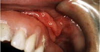
Patient presented to you after fitting the immediate denture 5 – 10 months, complaining pain and over tissue in the mandibular, what is the diagnosis:
1- Epulis fissurment.***
2- Hypertrophic Frenum.
------------------------
Fissured epulis (EF), a fissured tumor due to a traumatogenic prosthesis or fibrous inflammatory hyperplasia, is a hyperplastic growth of the mucosa in the gum or vestibular groove, in relation to the edge of a denture that gives it a cleft or cracked appearance.
Ethipatogenesis:
Fissured epulis results from chronic irritation for a long time, from the edge of a poorly adjusted denture so that the prosthetic mismatch causes a resorption of the supporting alveolar bone, which allows the denture to move and settle further down.
Clinical features:
The characteristic cracked epulis is formed by an elongated soft tissue impeller, which is initially red in color, is located in the vestibular course where the edge of the denture contours which in turn forms the fissure or ditch that determines its name ; the lesion may be located in the upper or lower gum; when it takes a long time to evolve it becomes paler than the adjacent mucosa, it may be ulcerated in the fissure.
Pathological anatomy:
The fissured epulis forms a mass of well-defined tissue of the normal mucosa, but not encapsulated, according to its evolution may have vascularized appearance or dense fibrous tissue. Microscopically it is constituted by hyperplastic fibrous tissue, with endothelial proliferation, the recent ones have good vascularization, on the contrary those of the long evolution have few vessels and abundant fibroblasts and collagen fibers, the epithelium that covers the fibrous mass can present acanthosis, inflammation Chronic rounded cell is more prominent near; the fissure
The prognosis of fissured epulis is good after a limited exeresis, it is considered a preneoplastic lesion, we have seen cases of malignization of the epithelium subjected to constant long-term trauma.
Types of epulis:
- Fibrous epulis.
- Granulomatous epulis.
- Fibrous epulis:
Fibrous epulis, considered by some authors as an irritating fibroma, is a formation of fibrous tissue, a fibrous hyperplasia resulting from chronic irritation that grows in a limited way at any site of the oral mucosa.
Causes:
The cause of fibrous epulis is similar to that of fissured epulis, it consists of a reaction of the oral tissues to chronic long-term trauma, but its development mechanism is not limited to the relationship with the edge of a denture, but it can be seen in various sites of the mucosa of the mouth and respond to different irritative factors.
Clinical features:
Fibrous epulis can grow on any site of the oral mucosa, but its preferred site is the gum, both upper and lower, its shape is rounded, it is attached to the underlying tissue by a sessile or pedicle base, its coloration is paler than the surrounding mucosa and its consistency is firm, when it is located in the gum, near the teeth it seems to originate in the periodontal ligament tissue.
Pathological anatomy:
It is a well-defined formation of a homogeneous surface and of a fibrous aspect, it is not very vascularized, when it is long-standing it can have calcified foci in its central part.
Microscopically, bands of dense connective tissue are observed, with few fibroblasts and scarce blood vessels, the stratified epithelium that covers it may be spherical and slightly hyperkeratotic. The prognosis is similar to that of fissured epulis.
- Granulomatous epulis:
Granulomatous epulis or gingival granuloma is a localized reaction to an irritation or infection, characterized by a rapid and exuberant formation of granulation tissue.
Causes:
Granulated epulis arises from the exaggerated proliferation of granulation tissue, as a tissue repair mechanism, the organization of granulation tissue is aborted by the continuous proliferation of endothelial cells stimulated by a foreign body such as a tooth fragment or a bone spicule that it remains in the socket after a dental extraction, sometimes caused by fragments of amalgam that are traumatizing the gum, after a careless filling.
Clinical features:
Granulomatous epulis grows in the form of a mass of dark red, bleeding, soft, painless tissue of usually small size from the edges of a traumatized wound or on the socket, after laborious extraction, it can be accompanied by ulceration and suppuration; It can feel itchy, rarely reaching a considerable size.
Pathological anatomy:
The tissue mass of this formation is soft, bleeding in appearance and precise limits, at its center it is possible to find the foreign body causing the exuberant proliferation.
Microscopically it is formed by a prominent proliferation of endothelial cells that exceed fibroblastic activity, today a marked acute inflammatory infiltrate, sometimes these lesions, when they are not surgically removed, suffer a fibrosis process over time, when the stimulation of the cells ceases endothelial, and become a fibrous epulis.
The prognosis of granulomatous epulis is magnificent, but to prevent recurrence, after its elimination, it is necessary to eliminate the foreign body causing proliferation, it is not a preneoplastic entity.
